CARL ZEISS MICROSCOPY EDUCATION AND RESEARCH CENTER
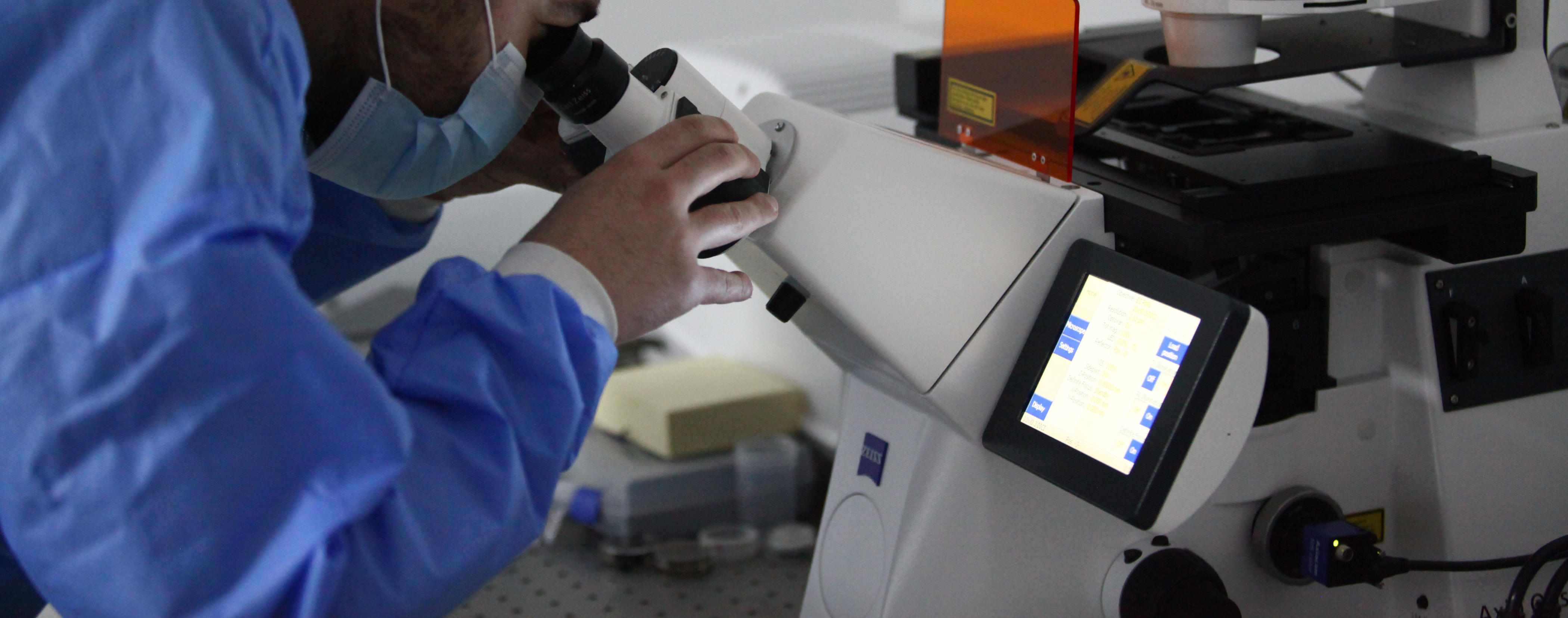
Carl Zeiss Microscopy Education and Research Center at New Vision University is a state-of-the-art facility equipped with modern photonic and electron microscopy technologies. As the only center of its kind in the region, it serves as a multidisciplinary platform for advanced research, scientific innovation, academic training, and clinical applications. Powered by cutting-edge instrumentation and an advanced live-cell volume light microscopy (4D) as well as volume electron microscopy (3D) imaging, the center is a focal point for collaboration in cell and tissue biology, micro- and nano-scale visualization technology, oncology, development of new biomaterials, new chemotherapeutic drugs research and development, neuroscience, and beyond.
The center welcomes national and international partnerships across academia, research, healthcare, and biotechnology. The open-access model enables shared use of instrumentation and expertise. Whether you are a student, researcher, or industry partner, our center offers a dynamic environment to explore, analyze, and innovate.
Our Mission
· Empower Education through hands-on microscopy training and interdisciplinary learning.
· Support Research with cutting-edge imaging technologies and expert guidance.
· Foster Innovation by enabling collaborations between academia, industry, and clinical institutions.
We aim to be a catalyst for scientific discovery and a bridge between emerging technologies and real-world applications.
Our Vision
To become a leading center of excellence in microscopy-driven biomedical research, where education meets innovation and where students, scientists, and clinicians converge to explore the unseen world at tissue, cellular, subcellular and molecular levels.
Scientific Infrastructure
|
Our center is equipped with high-performance instruments, including:
|
 |
 |
We also maintain an interesting cell culture lines including:
|
To ensure sustainable excellence, the Center relies on a continuous supply of essential reagents, labware, CO₂, and liquid nitrogen; Fully designed and properly zoned laboratory spaces for clean work, imaging, and cell culture; and maintenance of advanced infrastructure, including 3D/4D imaging software like Amira and Imaris, along with high-performance computing systems.